David Spears

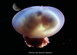
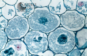
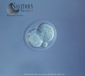
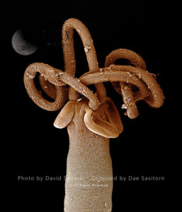
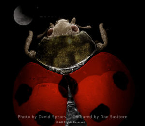
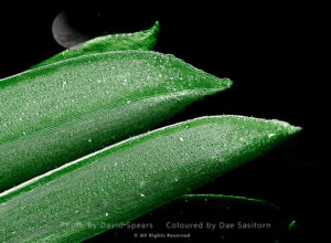
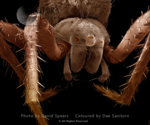
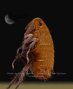
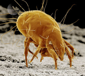
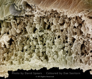
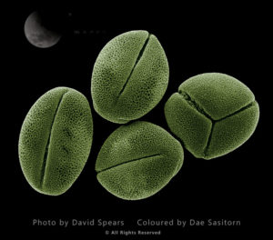
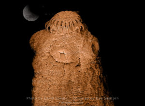
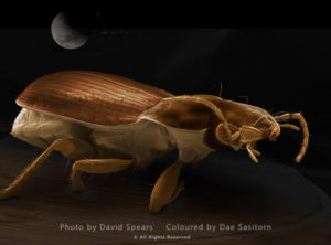
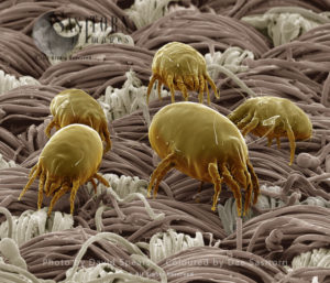
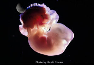
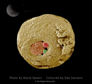
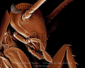
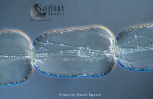
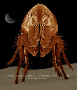
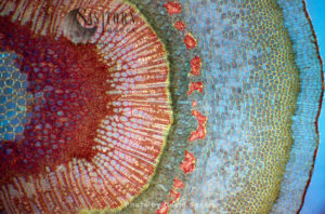
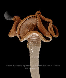
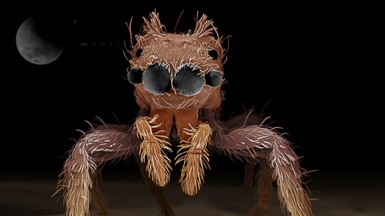
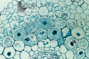
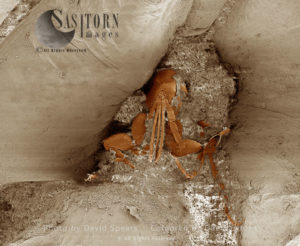
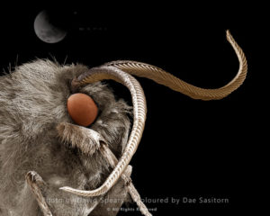
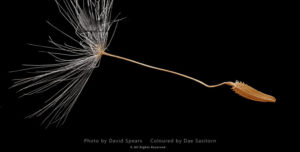
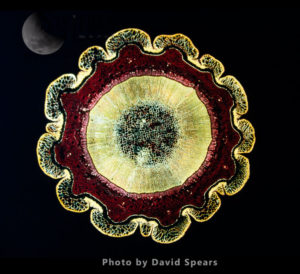
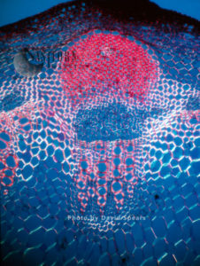
Photography: Macro and Micro Photography

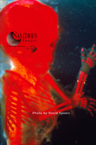
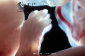
David Spears is a retired cameraman, producer and photographer. He is a Zoologist graduate from London University and holds two FRPS distinctions, an ASIS qualification from the RPS and is a Fellow of the Royal Microscopical Society.

His companies Science Pictures and Clouds Hill Imaging produced over 300 documentary programmes for BBC, C4, ITV, Discovery etc. Some won awards from Royal Television Society, British medical Association, and European Educational Association among others.

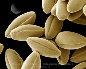
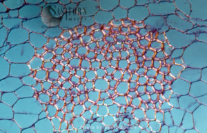
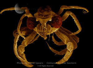
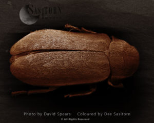
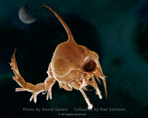
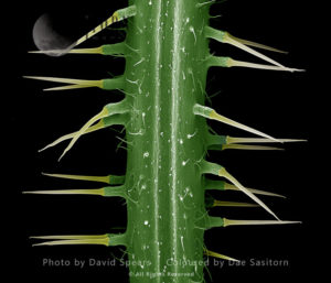
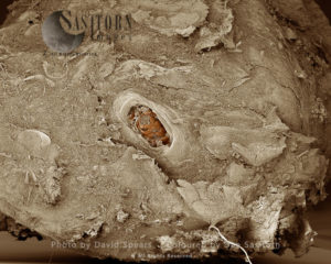
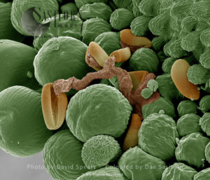
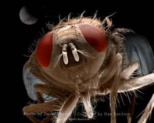
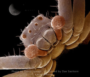
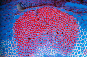
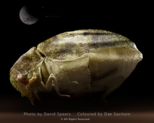
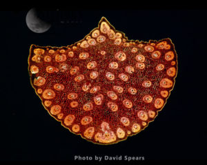
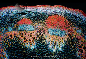
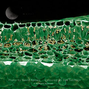
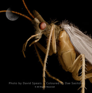
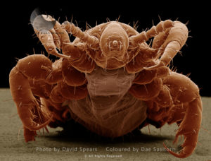
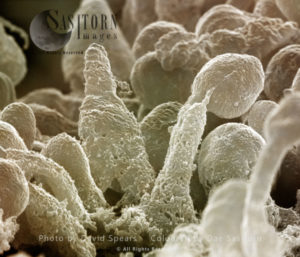
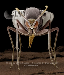
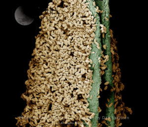
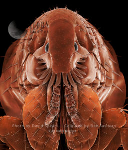
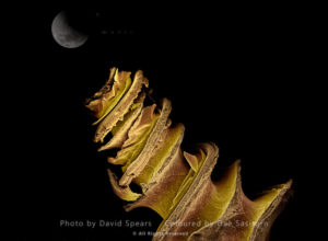
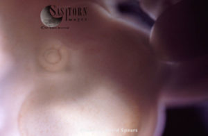
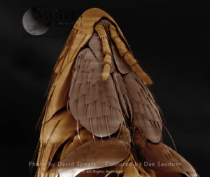
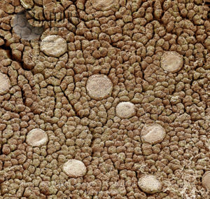
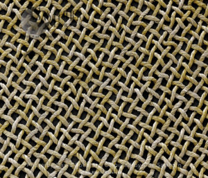
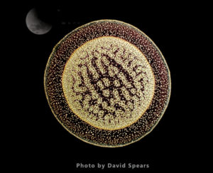
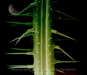
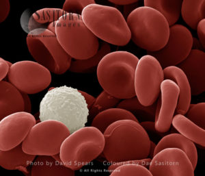
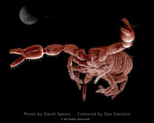
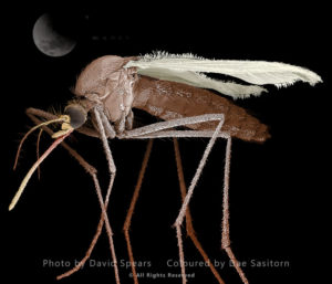
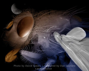
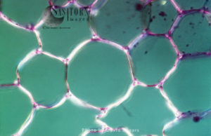
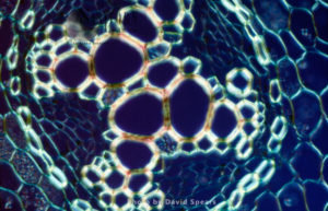
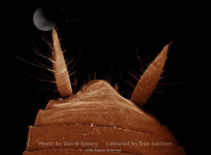
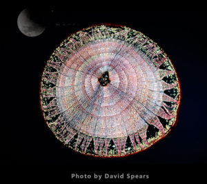
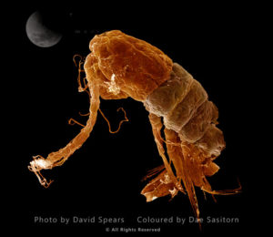
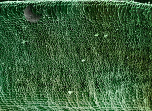
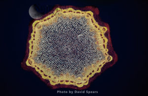
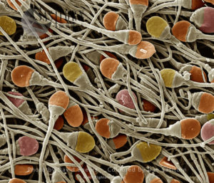
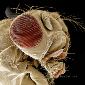
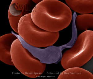
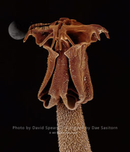
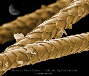
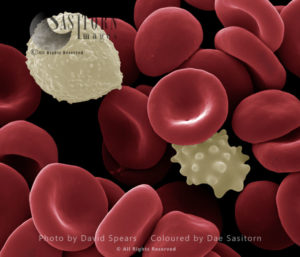
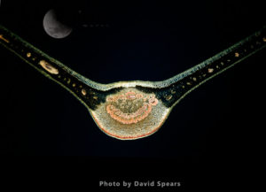
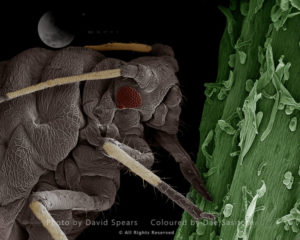
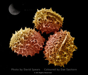
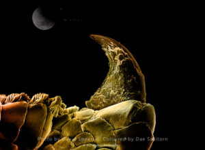
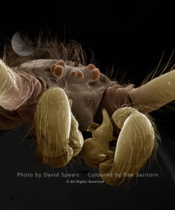
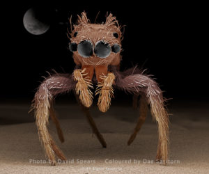
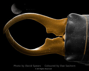
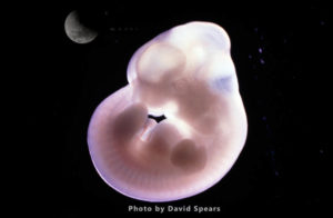
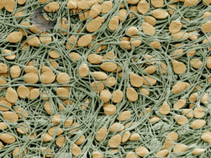
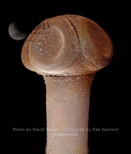
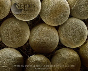
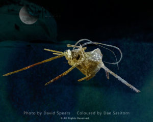
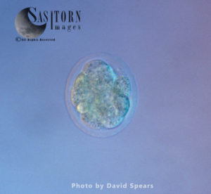
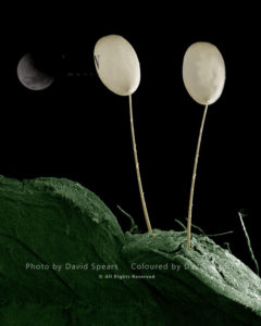
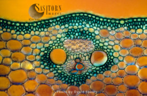
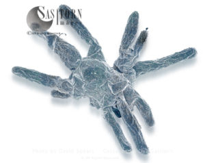
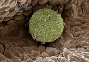
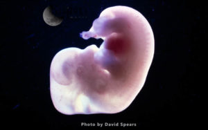
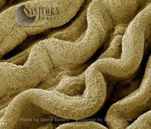
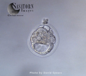
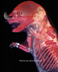
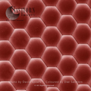
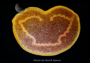
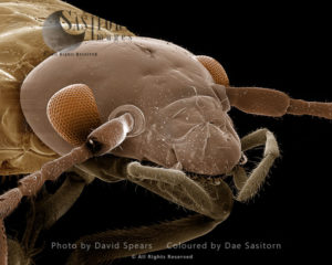
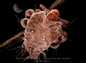
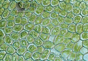
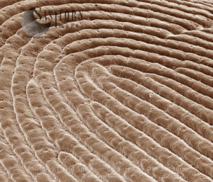
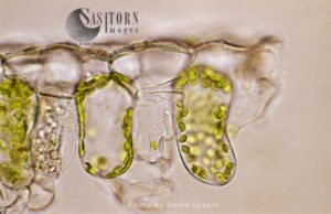
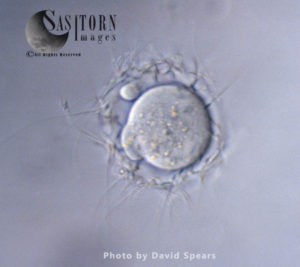
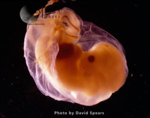
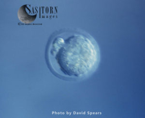
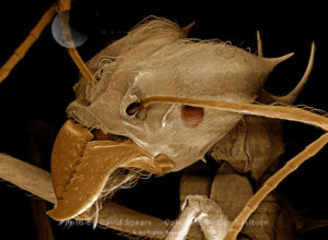
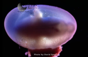
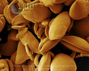
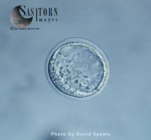
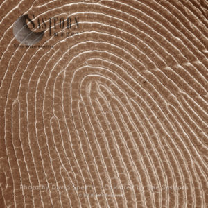
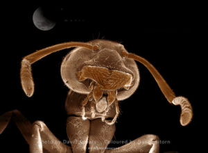
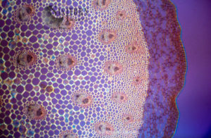
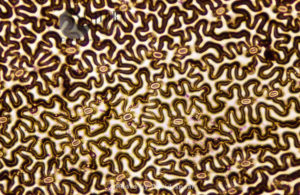
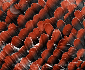
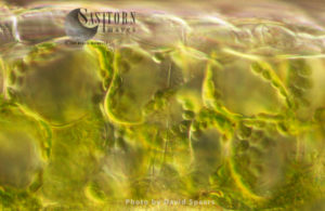
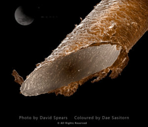
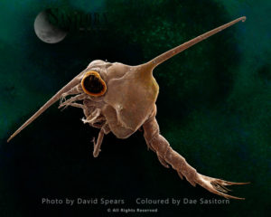
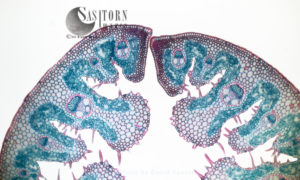
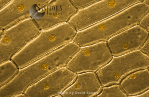
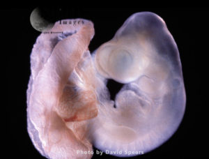
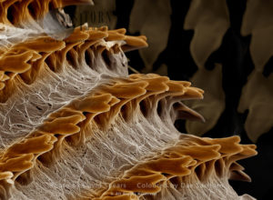
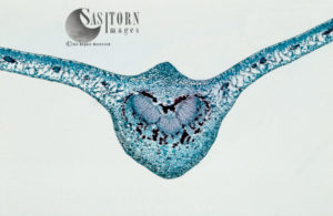
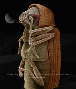
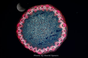
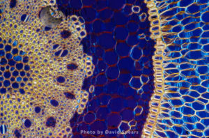
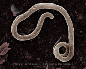
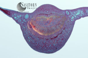
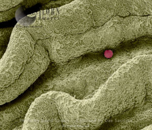
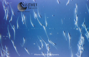
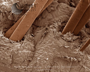
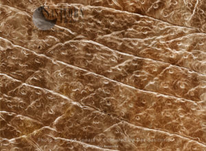
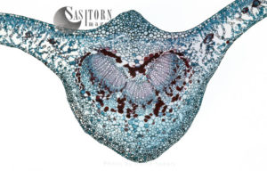
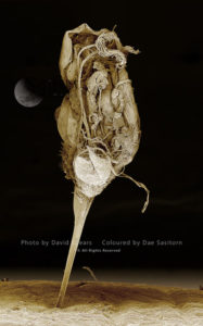
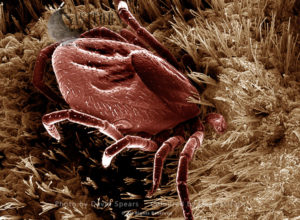
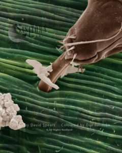
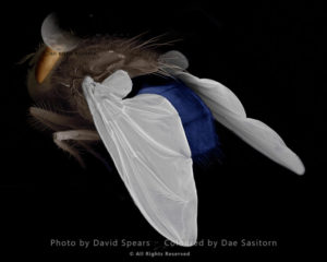
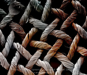
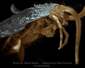
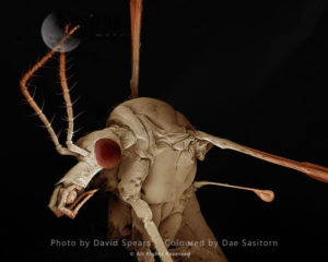
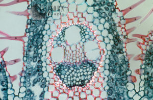
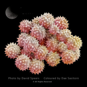
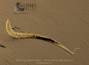
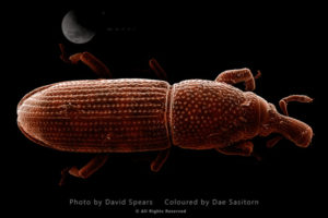
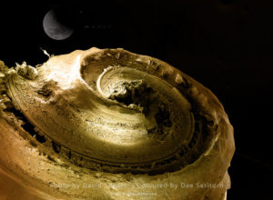
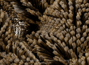
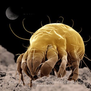
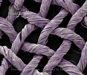
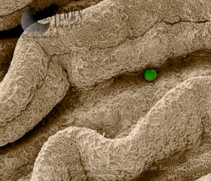
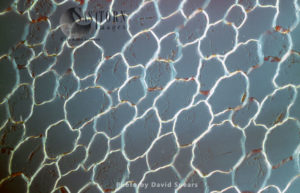
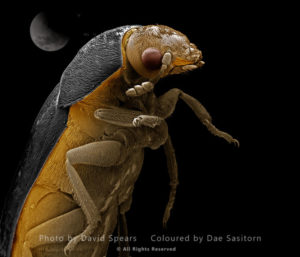
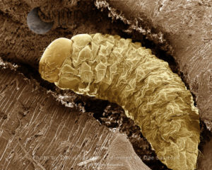
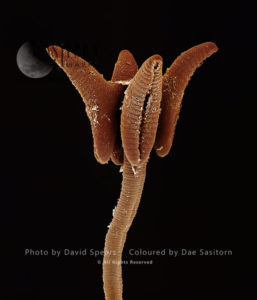
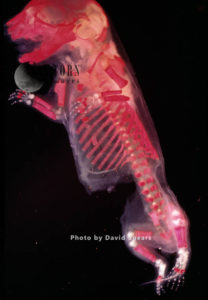
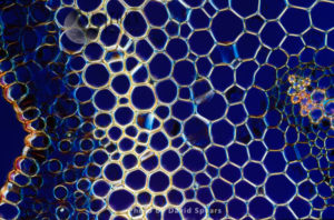
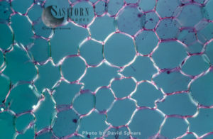
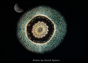
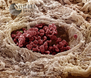
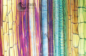
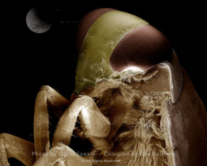
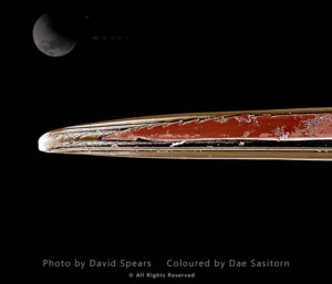
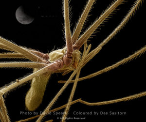
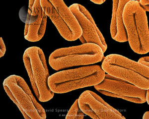
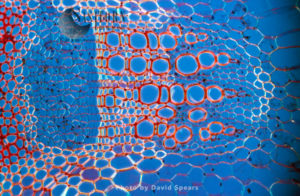
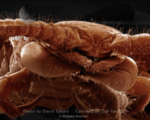
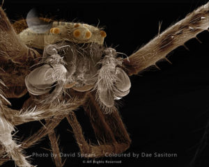
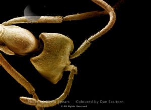
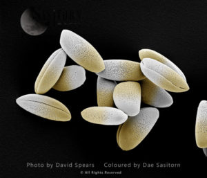
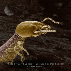
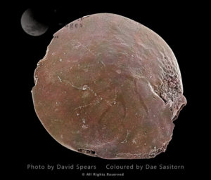
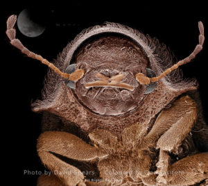
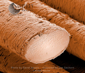
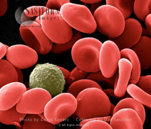
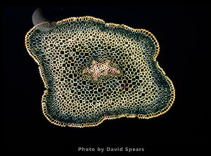
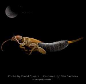
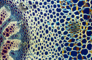
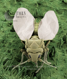
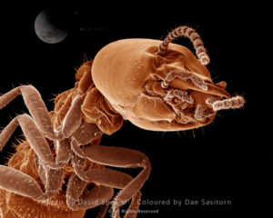
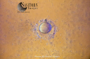
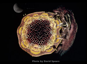
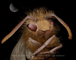
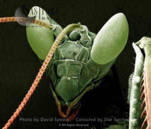
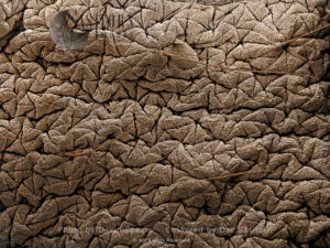
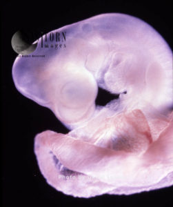
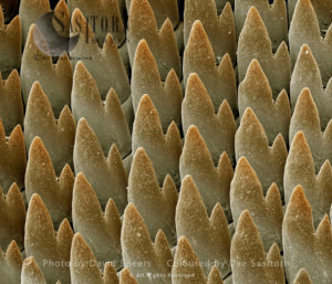
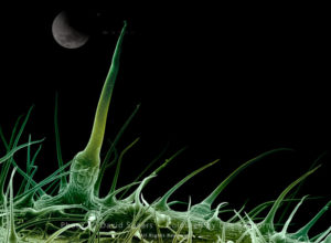
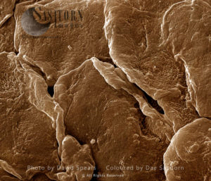
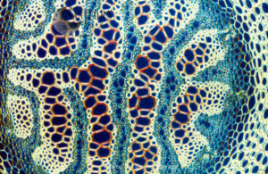
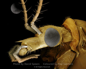
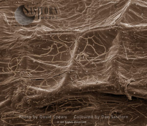
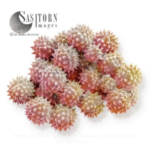
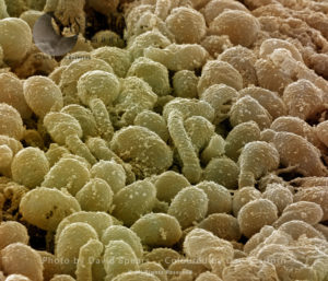
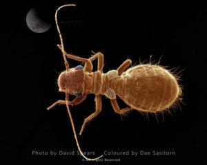
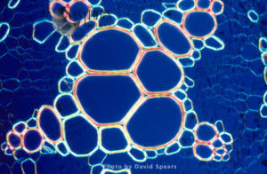
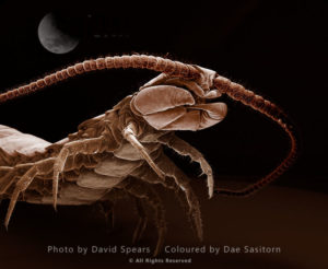
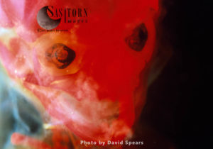
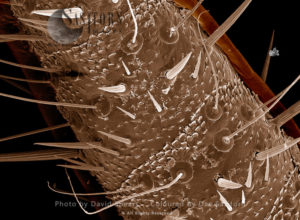
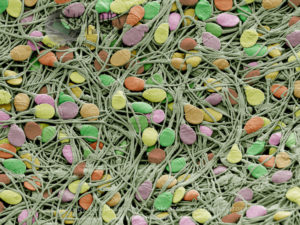
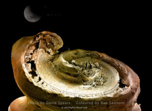
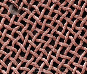
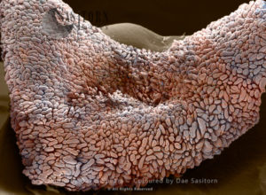
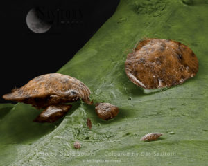
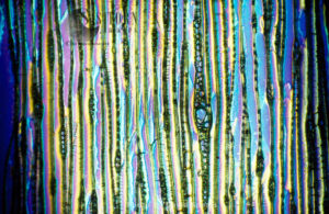
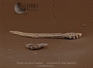
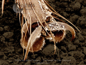
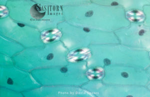
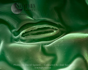
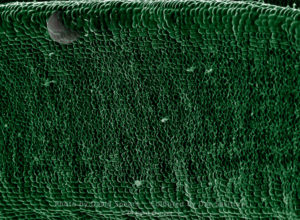
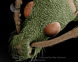
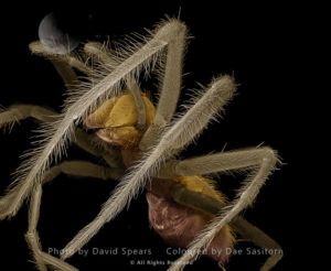
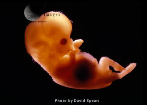
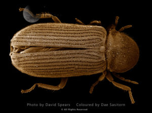
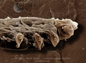
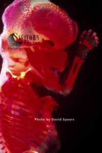
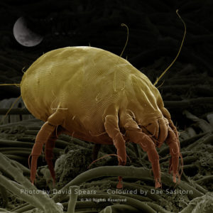
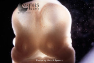
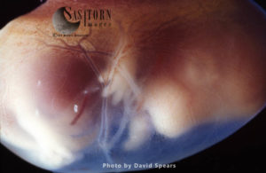
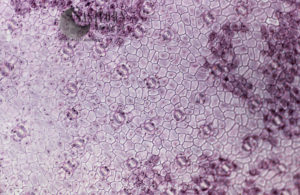
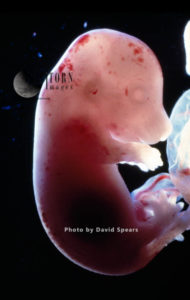
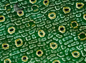
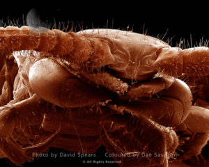
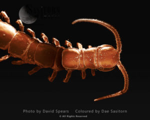
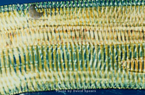
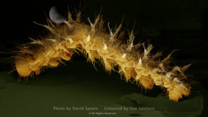
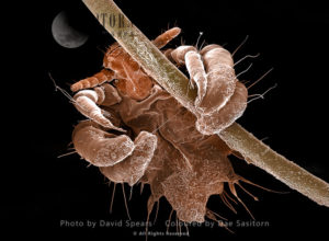
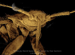
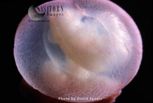
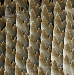
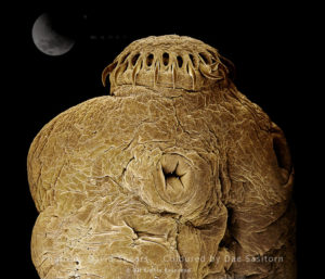
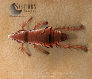
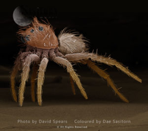
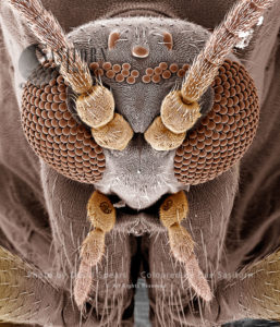
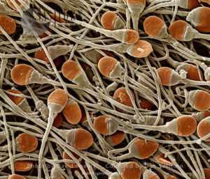
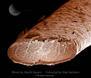
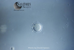
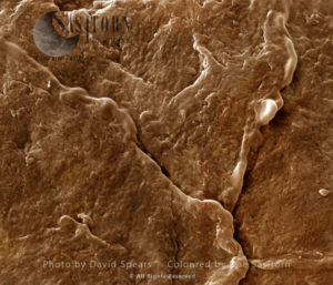
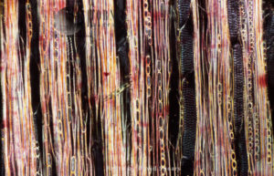
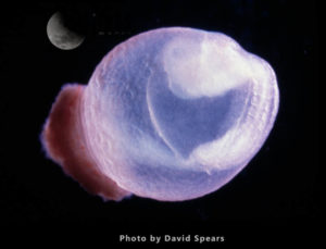
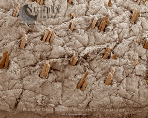
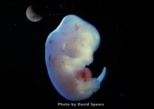
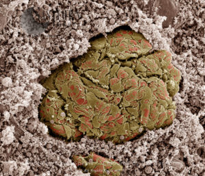
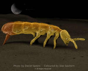
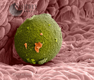
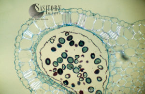
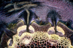
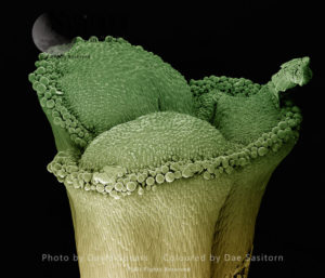
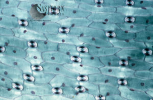
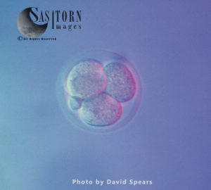
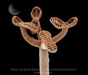
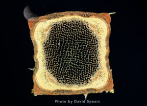
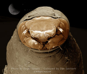
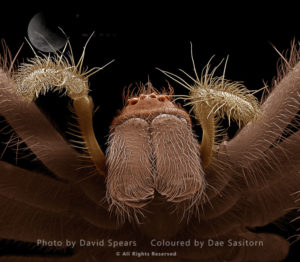
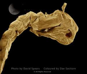
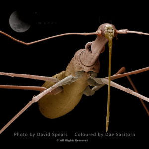
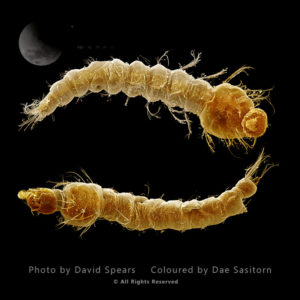
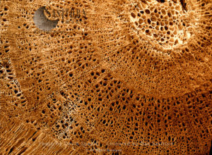
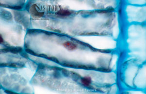
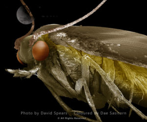
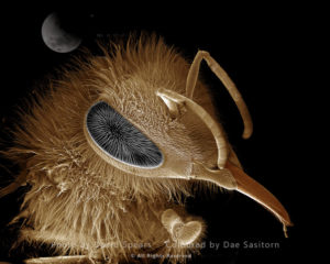
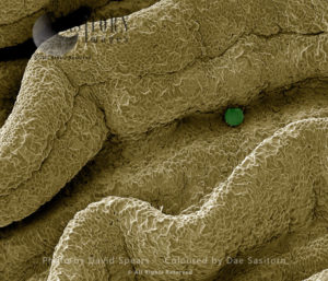
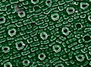
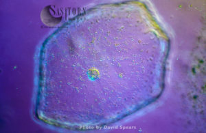
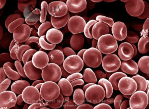
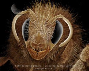
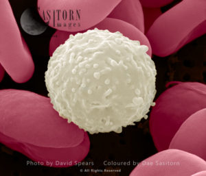
David delights in exploring the range of imaging techniques used by scientists and technologists and provides images that demonstrate that informative pictures produced for scientists do not have to be unattractive and badly composed.

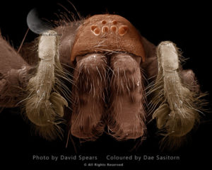
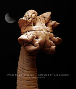
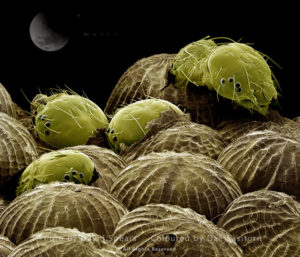
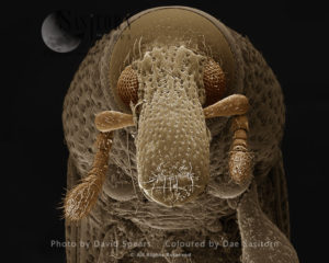
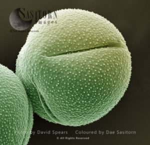
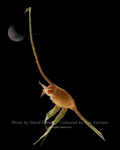
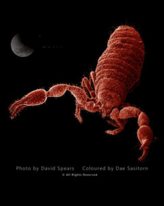
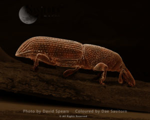
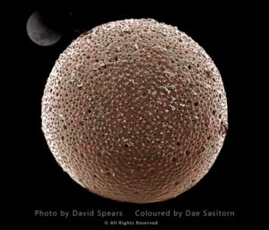
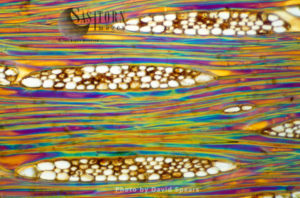
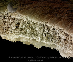
The equipment in his studio includes 1967 Zeiss photomicroscope converted to LED lighting and mirror less digital cameras and a Zeiss LEO 430 scanning electron microscope. David has also been pioneering the focus stacking technology, building two different systems, working with mirror less cameras.

Published book: Unseen Companions: Big Views of Tiny Creatures (published by Last Refuge)

Light Micrograph (LM): transverse section shows Sclerenchyma Ground Tissue in Helianthus stem
Megalop
Death Watch Beetle
Crab larvae
Chigoe Flea
Germination in Lilly
House Fly
Crab Spider
Light Micrograph (LM): Transverse section shows Sclerenchyma Ground Tissue in Helianthus stem
Varied Carpet Beetle
Light Micrograph (LM): A transverse section of a stem showing vascular bundle, cortex and epidermis of Jerusalem artichoke (Helianthus tuberosus)
Leaf Section
Caddis Fly
Human Head Louse
Human Retina
Fungus Gnat
anther of a lily with pollen
Cat Flea
Cochlea of Guinea Pig
Rat Embryo
Green Lacewing
Oriental Rat Flea
Frog tongue
Scanning Electron Micrograph (SEM): Silk fabric material
Light Micrograph (LM): A transverse section of an aerial root from a Pandanus sp
SEM: Stinging Nettle, Urtica dioica
Human White and Red Blood Cells
False Scorpion
Mosquito, Culex pipiens
Bluebottle
Rat Embryo
Light Micrograph (LM): Filaments of fungi Endotrophic Mycorrhiza live within cells of a root
Two cell stage mouse embryo
Tapeworm from shark
Light Micrograph (LM): transverse section shows Parenchyma or packing tissue from Cucurbita Stem
Light Micrograph (LM): transverse section showing xylem of root of Ranunculus repens (Creeping Buttercup)
American Cockroach
Light Micrograph (LM): A transverse section of a stem of Common Lime tree (Tilia europaea)
a gut tumour
Seven-Spot Ladybird
Yew Leaf
Garden Spider-female
Oriental Rat Flea
Calanus
Scanning Electron micrograph (SEM): Yew (Taxus baccata) Leaf
Light Micrograph (LM): A transverse section of a Dandelion stem (Taraxacum officinale)
Human sperm
Fruit Fly
Sleeping Sickness Parasite in red blood cells
Tapeworm from a blue shark
Cat Hairs
Light Micrograph (LM): A longitudinal section showing xylem elements of a Ribes sp. stem
Human White and Red Blood Cells
Light Micrograph (LM): A transverse section of a Fig leaf
Black Aphid
Gecko Claw
Pholcus Spider - male
A human Ovum  being fertilized
Jumping Spider
Earwig
Rat Embryo
Human sperm
Cheese Mite
Human Retina
Scanning Electron Micrograph (SEM): Peony Pollen
Tapeworm, Taenia solium
Tapeworm from a teleost fish
Spider Eggs
Dinoflagellate, Ceratium
Eight cell stage mouse embryo
Eggs of Green Lacewing
Light Micrograph (LM): A transverse section of a Maize stem (Zea sp.) showing vascular bundle, cortex and epidermis
Amoeba
SEM: Human Ovum in Fallopian Tube : Magnification x 1,000 for A4 size print
Ground Beetle
Dust Mites
Rat Embryo
Human embryo
Rat Embryo
Human Fallopian Tube
A Mouse Blastocyst
Rat Embryo
Eye facets of a House Fly
Lily Pollen
Light Micrograph (LM): A transverse section of a stem of a Tree Fern (Dicksonia antractica)
Earwig
Human Crab Louse
Light Micrograph (LM): The cellular struture of a non-vascular plant: Liverwort (Hepatica) showing chloroplasts and oil bodies
Human finger tip print
Light Micrograph (LM): plant cell chloroplasts - the site where photosynthesis takes place
A Mouse Ovum being fertilized
Human Embryo
Single cell stage mouse embryo
Leaf Cutter Ant
Rat Embryo
A Mouse Blastocyst
Human finger tip print
Gecko Claw
Black Garden Ant
Light Micrograph (LM): A transverse section of a stem of a Butcher's broom (Ruscus aculeatus)
Light Micrograph (LM): A concentration of cells on the epidermis of a plant showing stomata
Red Admiral Butterfly’s Wing Scales
Light Micrograph (LM): plant cell chloroplasts - the site where photosynthesis takes place
Human Hair - African
Crab larvae
Light Micrograph (LM): Transverse section of a leaf of Marram Grass (Ammophila sp.)
Light Micrograph (LM): Onion skin cells
Rat Embryo
Driver Ant
Light Micrograph (LM): transverse section shows Sclerenchyma Ground Tissue in Helianthus stem
American Cockroach
Light Micrograph (LM): Cells from a hair on the stamen of the common spiderwort (Tradescantia)
Cat Flea
Light Micrograph (LM): A Tranverse section of a stem of a Common or European Ash tree (Fraxinus excelsior)
Rat Embryo
Light Micrograph (LM): transverse section of Prunus Leaf
Woodworm or Furniture Beetle
Light Micrograph (LM): A transverse section of a Helianthus stem
Light Micrograph (LM): A transverse section of an aerial root of Orchid (Dendrobium sp.)
Micro-filarial worm
Light Micrograph (LM): A transverse section of a leaf of Tasmanian Blue Gum (Eucalyptus globulus)
SEM: Human Ovum in Fallopian Tube : Magnification x 1,000 for A4 size print
Human Sperm
Human Skin with Hair Follicles
Human skin
Light Micrograph (LM): A transverse section of a leaf of a Cherry (Prunus sp.)
Bee Sting
Black-legged or Deer Tick
Black Aphid
Bluebottle
Scanning Electron Micrograph (SEM): Silk fabric material
Silverfish
Crane Fly or ‘Daddy-Long-Legs’
Light Micrograph (LM): Transverse section of a leaf of Marram Grass (Ammophila sp.) showing Vascular Bundle;\
Common House Spider - male
Tapeworm from shark
Spectacle Moth hatching
Groundsel Pollen
C elegans
Wood-boring Weevil
Human Cochlea
Rat Embryo
Light Micrograph (LM): transverse section shows Sclerenchyma Ground Tissue in Helianthus stem
Woodboring Weevil
SEM: Cactus Pollen
Tapeworm from shark
Jumping Spider
Light Micrograph (LM): Filaments of fungi Endotrophic Mycorrhiza live within cells of a root
Chigoe Flea
Gecko foot hairs
Flour Mite
Scanning Electron Micrograph (SEM): Silk fabric material
SEM: Human Ovum in Fallopian Tube : Magnification x 1,000 for A4 size print
Light Micrograph (LM): transvers section shows Parenchyma Ground Tissue in Buttercup stem
Leather or Hide Beetle
Lavae of Woodworm or Furniture Beetle
Tapeworm from shark
Rat Embryo
Light Micrograph (LM): A transverse section of a stem of Whisk Fern (Psilotum nudum)
Light Micrograph (LM): tranverse section shows Parenchyma or packing tissue from Cucurbita Stem
Light Micrograph (LM): A transverse section of an aerial root of Orchid (Dendrobium sp.)
Crab larvae
False Scorpion or Pseudoscorpion
Wood-boring Weevil
Foraminifera
Human vein showing red blood cells
Light Micrograph (LM): Longitudinal section showing xylem elements of old sunflower stem(Helianthus sp.) showing spiral chloroplasts
Backswimmer
Wasp Sting
Pholcus Spider - female
Scanning Electron Micrograph (SEM): Pollen grains from an Umbelliferous flowering plant
Light Micrograph (LM): A transverse section shows Xylem in Helianthus stem
Common Centipede
Garden Spider-male
Driver Ant
Termite (woker)
Foraminifera
Deathwatch Beetle
Human Hairs - European
SEM: Human White and Red Blood Cells; Magnification x 21,000 for A4 size print
Light Micrograph (LM): A transverse section of a stem of Whisk Fern (Psilotum nudum)
Earwig
Light Micrograph (LM): A transverse section of a stem of Clubmoss(Lycopodium sp.)
Whitefly
Termite (woker)
A human Ovum  being fertilized
Light Micrograph (LM): A transverse section of a stem of Clover (Trifolium sp.) with an unidentified parasite attached.
Common Wasp
Praying Mantis
Human Skin
Rat Embryo
SEM: Stinging Nettle, Urtica dioica
Human skin
Light Micrograph (LM): A transverse section of a stem of Clubmoss(Lycopodium sp.)
Crane Fly or ‘Daddy-Long-Legs’
Bedbug
Human Hair - Asian
Human sperm fertilizing egg (ovum)
Sleeping Sickness Parasite in red blood cells
Scanning Electron Mirograph (SEM): Mycorrhizae Fungi on root
Groundsel Pollen
Human Retina
Psocid
Light Micrograph (LM): A transverse section showing xylem of a root of Creeping Buttercup, (Ranunculus repens)
Common Centipede
Human Foetus
American Cockroach
Footman moth
Dandelion seed
Light Micrograph (LM): The transverse section of a stem of Norway Pine (Abies Excelsa) shows the resin passages
Light Micrograph (LM): A transverse section shows Vascular Bundle in Helianthus stem
Human sperm
Human Cochlea
Scanning Electron Micrograph (SEM): Silk fabric material
Scanning Electron Micrograph (SEM): Human gut showing villi
Brown Scale Insect
Light Micrograph (LM): Longitudinal section showing xylem elements of Scots pine wood, (Pinus sylvestris)
Follicle Mite
A foot of a House Fly
Lily Pollen
SEM: Human White and Red Blood Cells; Magnification x 21,000 for A4 size print
Human Head Louse
Human testis tubes
Light Micrograph (LM): A transverse section of a leaf of a Tulip (Tulipa sp.) showing Stomata
Scanning Electron micrograph (SEM): Open Stoma of  Chrysanthemum plant leaf
Scanning Electron micrograph (SEM) showing stomata on a Yew (Taxus baccata) Leaf
Leaf Weevil
Common House Spider - female
Human Embryo
Powder Post Beetle, Priobium sp.
Follicle Mite
Human Foetus
Dust Mite
Rat Embryo
Rat Embryo
Light Micrograph (LM): A transverse section of a lower Epidermis of a Box leaf (Buxus sp.) showing Stomata
Rat Embryo
Scanning Electron micrograph (SEM) showing stomata on a Yew (Taxus baccata) Leaf
Common Centipede
Common Centipede
Light Micrograph (LM): Longitudinal section shows Scalariform vessels of Fern
Small Tortoiseshell Butterfly
Human Crab Louse
Bedbug
Rat Embryo
Tapeworm, Taenia solium
Light Micrograph (LM): Longitudinal section showing xylem elements of Mahogani (Swietenia mahagoni)
Human Retina
Cat Flea
Jumping Spider
Human Foetus
Rat Embryo
Fungus Gnat
Human sperm
Human Public Hair
A Mouse Ovum being fertilized
Human skin
Light Micrograph (LM): A longitudinal section showing xylem elements of a Ribes sp. stem
Rat Embryo
Human Skin with Hair Follicles
Rat Embryo
Kidney glomerulus
Springtail
SEM: Human Ovum in Fallopian Tube : Magnification x 1,000 for A4 size print
Light Micrograph (LM): Transverse section of Dehiscence Lilium Anthers with Pollen
Light Micrograph (LM): A transverse section of a stem of Clover (Trifolium sp.) with an unidentified parasite attached.
stigma of a lily
Light Micrograph (LM): A transverse section of a leaf of a Tulip (Tulipa sp.) showing Stomata
Four cell stage mouse embryo
Tapeworm from a batoid fish
Bluebottle
Human Sperm
SEM: Human Red Blood Cells; Magnification x 6,500  for A4 size print.
Black-legged or Deer Tick
Light Micrograph (LM): A transverse section of a stem of a Hedge Woundwort plant (Stachys sylvatica)
Crane Fly or ‘Daddy-Long-Legs’ lavae
Common House Spider - female
Anopheles Mosquito pupa
Water Measurer
Anopheles Mosquito larvae
Wood structure. Coloured scanning electron micrograph (SEM) of a cross-section through a twig from a hardwood tree.
Light Micrograph (LM): A light micrograph of the nuclei of plant cells
Common Clothes Moth
Honeybee
Human Ovum in Fallopian Tube
Scanning Electron micrograph (SEM) showing stomata on a Yew (Taxus baccata) Leaf
Light Micrograph (LM): Tomato seed
Human Red Blood Cells
Bumblebee
SEM: Human White and Red Blood Cells; Magnification x 21,000 for A4 size print
Shopping cart
There are no products in the cart!
Continue shopping